 |
|
|
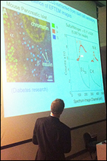
Caption: Richard Leapman, scientific director at NIH’s NIBIB, presents the latest uses of electron microscopes to discover the structure of the smallest parts of cells. |
|
Richard Leapman, scientific director of the National Institute of Biomedical Imaging and Bioengineering, a division of the National Institutes of Health, showed some new ways that biologists are using sophisticated electron microscopes to discover the workings of cells and their organelles at the most basic levels. They aim to gather nanoscale information about the architecture and composition of biological structures.
Leapman said that some of the advances include 3D images of thick structures that couldn’t have been thoroughly imaged before, examining individual atoms attached to biological structures, and examining the impact of human-built nanomedical particles on cells.
Leapman is Chief of NIBIB's Laboratory of Cellular Imaging and Macromolecular Biophysics, while also serving as NIBIB's Scientific Director since 2006.
April 10, 2014
|