|
Department of Mechanical Engineering faculty and graduate students have published new research in the Journal of the Mechanical Behavior of Biomedical Materials that could shed light on the mechanical cause underlying mild traumatic brain injuries (mTBI).
Recent media coverage of athletes and head injuries has drawn attention to the complex, and often puzzling nature of head injuries and the long-term damage that both severe and seemingly mild injuries can inflict on an individual.
While there is a large body of research being done on head injuries, especially, in the area of long-term cognitive impacts as a result of repeated head injuries, researchers at the University of Maryland are taking a closer look at the brain's mechanics, and how even seemingly minor trauma could cause damage, depending on how stress waves move through brain tissue due to its nature and composition and its ability to handle stress loads.
Mechanical Engineering research faculty member Dr. Henry Haslach and graduate student Lauren Leahy have collaborated with colleagues Peter Riley, a mechanical engineering alumnus and third-year medical student at the University of Maryland Medical School, Adam H. Hsieh from the University of Maryland’s Fischell Department of Bioengineering and Rao Gullapalli and Su Xu from the University of Maryland School of Medicine’s Department of Diagnostic Radiology and Nuclear Medicine on their paper, “Solid-extracellular fluid interaction and damage in the mechanical response of rat brain tissue under confined compression.”
 |
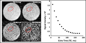
| The largest changes in the [brain] tissue after compression, compared to the state prior to compression, are observed near the plunger and near the base, even though the viscoelastic tissue relaxed during the time required for imaging. Read more. |
The aim of their research is to analyze the mechanical processes that might underlie mild traumatic brain injuries. According to the paper, “subtle small-scale mechanical damage mechanisms in brain tissue that can modify brain function may be involved in the in the initial cause of mTBI.” The brain is comprised of both solid matter and water, with a high fluid content, and the configuration of “brain tissue is maintained by the interaction between the cellular solid matter and the brain extracellular fluid (ECF).”
Based on this relationship, the team hypothesized that head trauma, even moderate strain, could cause stresses that could alter the brain’s mechanical properties, and their research results suggest "that trauma-induced increases in ECF hydrostatic pressure, which may induce pathological ECF flow, are a possible immediate mechanical cause of brain damage." Brain tissue, unlike cartilage or arterial tissue, is not a load bearing tissue, and the research indicates that when the brain is subjected to load bearing stress, such as that caused during head trauma, it experiences a loss in its stress-carrying ability, possibly due to cell rupture, disassociation of axonal-glial interconnections, relative motion of substructures in the heterogeneous tissue, or relative motion at interfaces or larger regions as a result of excessive hydrostatic pressure in the ECF.
While brain tissue is not particularly adept at handling repeated stress loads like other soft tissue, it does share soft tissue's property of propagating stress waves in a nonlinear fashion. This nonlinear property was what Department of Mechanical Engineering Chair and Minta Martin Professor of Engineering Dr. Balakumar Balachandran and doctoral student Marcelo Valdez took a closer look at in their publication, entitled "Longitudinal nonlinear wave propagation through soft tissue."
Through their combined analytical and numerical efforts, they analyzed the nonlinear effects of soft tissue mechanical behavior on propagating stress waves to create models that could be used for analyzing the effects of stress and impacts on soft tissue.
While the research was not aimed specifically at brain tissue, the results could be applied to work in both TBI and blast induced traumatic brain injuries, where an impact or an external blast, like an explosion, generates a load that is strong enough to create stress waves within the brain.
While many head injuries can often be attributed to the effects from motion between the brain and skull during impacts, there are few theories on what occurs in the brain during low impact injuries, or when there is an absence of motion between the skull and brain. Since even mild impacts are likely to cause stress waves in brain tissue, the researchers propose that better understanding of, and predictive modeling for, the behavior of these waves, including attenuation, speed, and frequency, could provide insights into the impacts of seemingly mild injuries on soft tissues such as the brain tissue.
A better understanding of the mechanical properties of brain tissue and how it handles stress from impacts could provide more clues into how even seemingly small head injuries can lead to greater or long term brain damage.
Both Drs. Balachandran and Haslach started their initial research efforts in this area through support from the Center for Energetic Concepts and Development.
For more information on the Haslach and his research, visit his faculty webpage.
For more information on Balachandran and his research, visit his faculty webpage.
Related Articles:
UMD Researchers Determine Process Behind Aortic Dissection
October 23, 2013
|

